Surgery. 2020 Feb 26. pii: S0039-6060(20)30051-9.
Authors
Kollar B, Rizzo NM, Borges TJ, Haug V, Abdulrazzak O, Kauke M, Safi AF, Lian CG, Marty FM, Rutherford AE, Mitchell RN, Murphy GF, Tullius SG, Riella LV, Pomahac B.
Abstract
BACKGROUND:
Long-term outcomes after face transplantation are rarely reported in the scientific literature. Here we present outcome data of a partial face allograft recipient 10 years after transplantation.
METHODS:
Medical records were reviewed for functional and psychosocial outcomes as well as complications. Histopathologic analyses of autopsy tissues and characterization of skin immune cells were performed.
RESULTS:
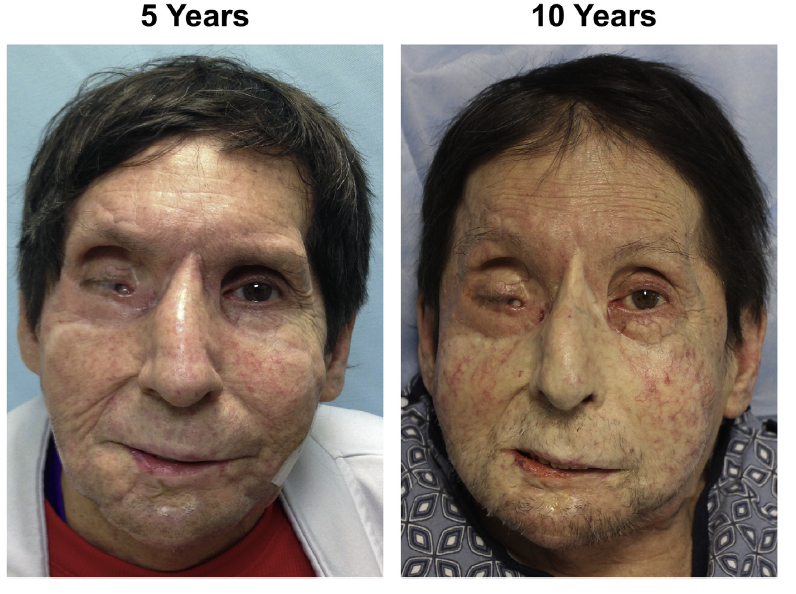
The patient retained long-term motor and sensory function, though with a noticeable drop in sensory function after year 5. Social reintegration of the patient was marked by reconnection with his family and participation in public social activities. Immunosuppressive therapy consisted of tacrolimus (target levels 6-8 ng/mL after the first year), mycophenolate, and prednisone, while steroids were completely weaned between years 1 and 7. One acute cellular rejection episode of grade II or higher occurred on average per year and led to chronic skin changes (papillary dermal sclerosis with superficial hyalinization, epidermal thinning with loss of rete ridges, perieccrine fibrosis), but the allograft vessels, muscles, adipose tissue, and bone were spared. Allograft skin was characterized by increased number of CD4+ TNF-α/IL17A producing T-cells as compared with native skin. Long-term kidney function was maintained at 60 mL/min estimated glomerular filtration rate. Unfortunately, the preexisting hepatitis C virus infection with liver cirrhosis was resistant to 3 treatments with new direct-acting antivirals and eventually hepatocellular carcinoma developed, causing the patient’s death 10 years after transplantation.
Figure. Aesthetic outcome of the patient. Patient is shown at the time of first histological signs of chronic skin changes (LEFT), and at maximal followup (RIGHT). At POY 10, the pale appearance of the allograft is pronounced as compared with the native skin. The patient provided written consent for publication of his photographs.